Hip Joint Loose Body Removal and Treatment in Bangalore
Writing
Loose bodies in the hip joint are small fragments of bone, cartilage, or other tissue that move freely within the joint. They may develop due to injury, degenerative joint changes, or certain joint conditions. These fragments can cause hip pain, stiffness, clicking, catching, or restricted movement, particularly when walking or rotating the hip. Loose bodies in hip joint treatment in Bangalore depends on the size and location of the fragments, the severity of symptoms, and the underlying cause. After a clinical examination and appropriate imaging, treatment may involve observation, activity modification, or hip arthroscopy to remove loose fragments when necessary.
Dr. Raghavendra Kembhavi can assess the condition and discuss suitable treatment options based on individual needs. The goal is simple: to remove the source of mechanical irritation and restore smooth, pain-free movement to your joint.
What Are Loose Bodies in the Hip Joint?
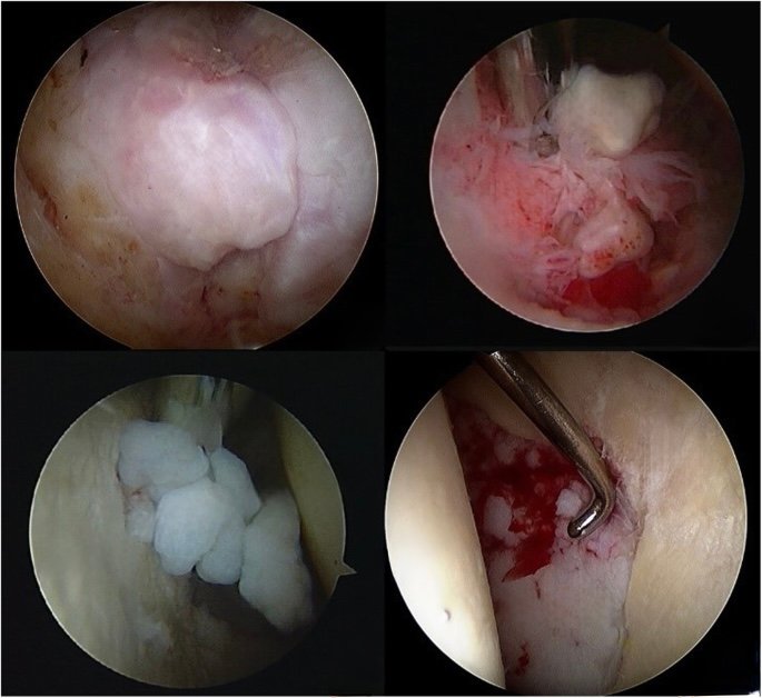
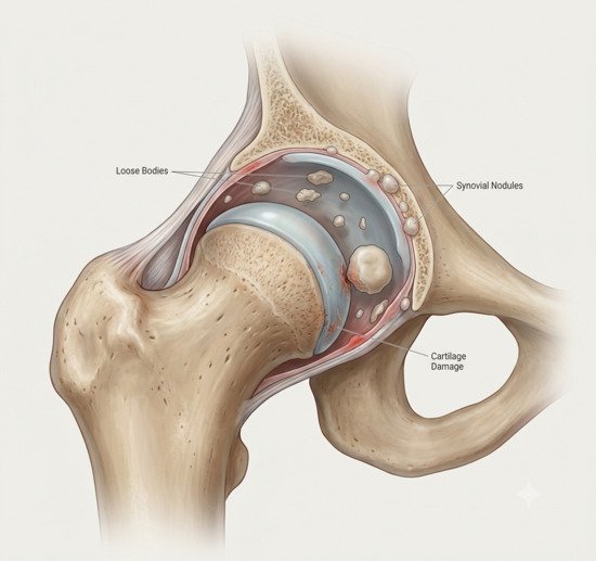
Loose bodies are exactly what they sound like — fragments of tissue that are floating freely within the joint. They can be made of bone, cartilage, or a combination of both. Some are the result of a single traumatic injury, while others develop gradually over time due to wear and tear or an underlying condition.
In the hip, loose bodies are often associated with conditions like synovial chondromatosis — a rare condition where the synovium produces multiple small cartilage nodules that can break off and float in the joint. They can also result from a previous injury, avascular necrosis, or degenerative changes. In the knee, they’re commonly seen after cartilage injuries, meniscal tears, or in conditions like osteoarthritis.
The problem with loose bodies is mechanical. They don’t belong there, and when they get caught between the moving surfaces of the joint, they cause pain, catching, and sometimes locking. Over time, they can also cause damage to the articular cartilage, leading to further joint deterioration.
Signs and Symptoms of Loose Bodies in the Hip Joint
Not everyone with loose bodies experiences symptoms. Some are discovered incidentally during imaging for another problem. When symptoms do occur, they’re usually mechanical in nature — meaning they’re related to movement rather than inflammation. Common signs include:
Catching or Locking
A sudden, sharp pain when the loose body gets caught between the joint surfaces. The joint may briefly lock or give way.
A Sensation of Something Moving
Many people describe feeling like something is floating or rolling inside the joint when they move it.
Intermittent Pain
Pain that comes and goes, often related to certain movements or positions. It may be sharp and fleeting rather than a constant ache.
Reduced Range of Motion
The joint may feel blocked or restricted, as if something is physically preventing it from moving through its full range.
Types of Loose Bodies
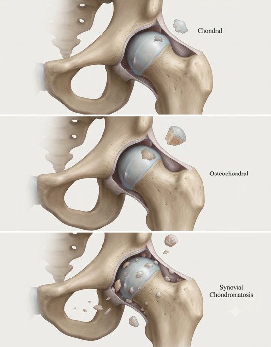
Loose bodies come in different forms, and understanding which type is present helps guide treatment. The main categories include:
Chondral Loose Bodies
These are fragments of pure cartilage that have broken off from the joint surface. They’re often seen after a traumatic injury or in cases of advanced cartilage degeneration. Because they’re made of cartilage, they may not be visible on standard X-rays.
Osteochondral Loose Bodies
These contain both bone and cartilage. They typically result from an injury that separates a piece of bone along with its overlying cartilage. Because they contain bone, they’re usually visible on X-rays and can be larger than pure chondral fragments.
Synovial Chondromatosis
This is a rare condition where the synovium itself produces multiple small cartilage nodules that can detach and float in the joint. Over time, these nodules can calcify and become numerous, causing significant mechanical symptoms and joint damage if not treated.
Diagnosis
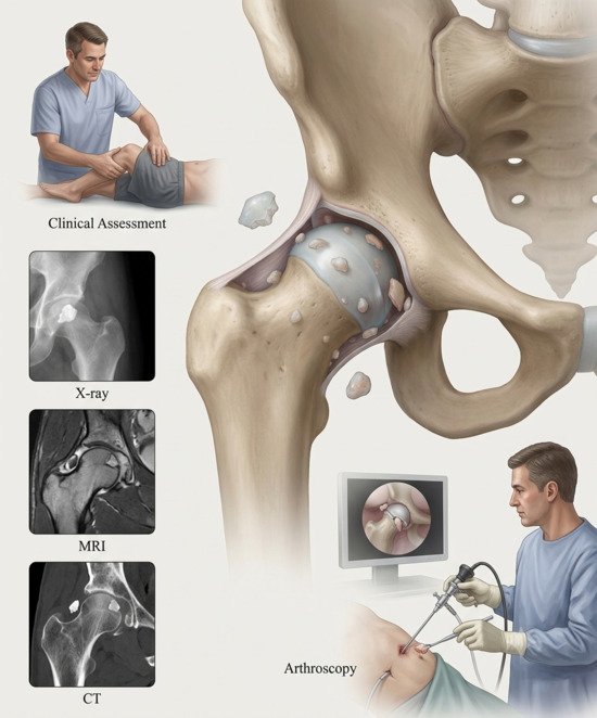
Diagnosing loose bodies requires a combination of clinical assessment and imaging. Dr. Raghavendra Kembhavi begins with a detailed history — when did the symptoms start, what movements trigger them, and is there any history of injury or previous joint problems? A physical examination assesses the joint for range of motion, catching, and any palpable fragments.
Imaging Studies
X-rays are often the first investigation. They can detect calcified loose bodies — those containing bone — but may miss purely cartilaginous fragments. An MRI is more sensitive and can identify both bony and cartilaginous loose bodies, as well as any associated damage to the labrum, cartilage, or ligaments. In some cases, a CT scan may be used to get a more detailed view of the bony anatomy and the exact location of the loose bodies.
Treatment Approach
The treatment of loose bodies depends on several factors: the size and number of fragments, the severity of symptoms, and whether there’s any underlying joint damage. Small, asymptomatic loose bodies may not require treatment and can simply be monitored. However, if they’re causing mechanical symptoms — catching, locking, or pain — removal is usually recommended.
Arthroscopic Removal
The gold standard for treating symptomatic loose bodies is arthroscopic removal. This minimally invasive procedure allows Dr. Kembhavi to visualise the joint directly, identify all loose fragments, and remove them through small keyhole incisions. In the same procedure, he can also address any underlying problems — such as a labral tear, cartilage damage, or synovial chondromatosis — that may have contributed to the formation of the loose bodies. Because it’s arthroscopic, recovery is faster and less painful than open surgery.
What to expect after surgery
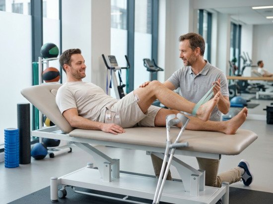
Recovery from arthroscopic loose body removal is generally straightforward. Because the procedure is minimally invasive, most patients experience less pain and a quicker return to function compared to open surgery. You’ll typically use crutches for a few days to a week, depending on the extent of the procedure and any additional work performed.
Physical therapy begins early to restore range of motion and strength. Most people can return to desk work within 1 to 2 weeks, while a full return to sports or heavy activity may take 4 to 8 weeks. If additional procedures were performed — such as a labral repair or cartilage treatment — the recovery timeline may be longer. Dr. Kembhavi will provide a personalised rehabilitation plan based on your specific situation.
Why choose Dr. Raghavendra Kembhavi
A focus on joint preservation
Dr. Kembhavi’s approach is built around preserving your natural joint. Rather than rushing to joint replacement, he explores every avenue to restore function and reduce pain through minimally invasive techniques.
Advanced arthroscopic skills
With fellowship training in arthroscopy and sports medicine, Dr. Kembhavi is experienced in the latest keyhole techniques for the hip, knee, and shoulder. This means smaller incisions, less pain, and faster recovery for his patients.
Personalised, patient-first care
Every patient is different, and so is every joint. Dr. Kembhavi takes the time to understand your specific situation — your goals, your lifestyle, and your concerns — and builds a treatment plan that’s tailored to you, not just your diagnosis.
Comprehensive diagnostic approach
Loose bodies are often a sign of an underlying problem. Dr. Kembhavi’s thorough diagnostic process ensures that the root cause is identified and addressed, not just the fragments themselves.