Cartilage Injuries Treatment in Bangalore
Cartilage injuries are often silent at first. There’s no dramatic pop like a ligament tear, no immediate swelling — just a gradual ache, some stiffness, and a feeling that your joint doesn’t quite move the way it used to. Over time, the pain becomes more persistent, and simple activities like climbing stairs or getting up from a chair start to feel difficult.
Articular cartilage is the smooth, slippery tissue that covers the ends of your bones. Unlike other tissues, it has no blood supply, which means it has very limited ability to heal on its own. When it’s damaged, the joint loses its protective cushion, and the bones begin to rub together. At our clinic in Bangalore, Dr. Raghavendra Kembhavi specialises in cartilage restoration procedures designed to preserve your natural joint and prevent or delay the need for replacement surgery.
What causes cartilage injuries?
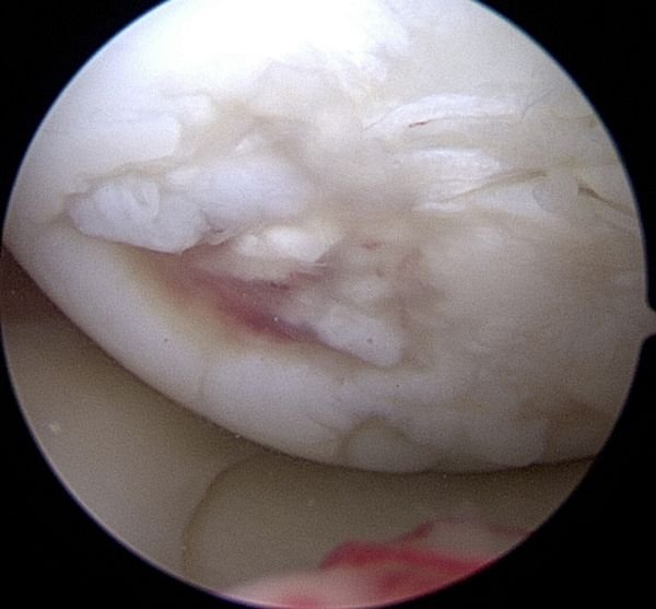
Articular cartilage is the smooth, white tissue covering the ends of bones in a joint. It allows bones to glide over each other with minimal friction and absorbs shock during movement. Because it has no blood supply, it cannot repair itself effectively — which is why even small defects can progressively worsen over time.
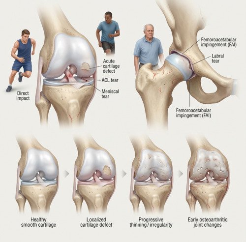
Cartilage injuries fall into two main categories. Acute injuries happen from a single traumatic event — a fall, a sports injury, or a direct blow that shears off a piece of cartilage. These are more common in younger, active people. Degenerative injuries develop gradually over years from repetitive wear and tear, often related to alignment issues, previous injuries, or simply ageing.
In the knee, cartilage damage often accompanies ligament injuries (ACL tears) or meniscal tears. In the hip, it’s frequently linked to femoroacetabular impingement (FAI) or labral tears. Wherever it occurs, the key is to address it before it progresses to osteoarthritis.
Signs and symptoms
Cartilage damage can be tricky to recognise because the symptoms come on gradually and are often dismissed as a minor strain. The pattern usually points to the diagnosis.
Persistent joint pain
A deep, aching pain that worsens with activity and may improve with rest. Often felt along the joint line.
Swelling and stiffness
Recurring swelling after activity and a feeling of tightness or reduced flexibility in the joint.
Grinding or catching
A sensation of grinding, popping, or catching during movement. In severe cases, the joint may lock.
Difficulty with stairs or squatting
Pain that intensifies when going up or down stairs, kneeling, or squatting — movements that load the joint.
Diagnosis
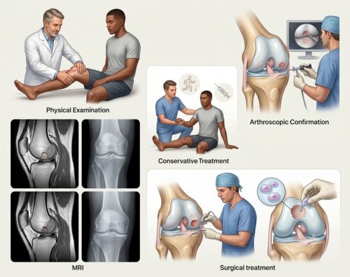
Diagnosis begins with a detailed history and physical examination. Dr. Kembhavi assesses your joint’s range of motion, stability, and areas of tenderness, and looks for signs of alignment problems that may be contributing to the cartilage damage.
Imaging and assessment
An MRI is the imaging study of choice for cartilage injuries. It shows the size, depth, and location of the defect, and helps determine whether the surrounding cartilage is healthy. X-rays can show joint space narrowing but don’t reveal cartilage directly. In some cases, a diagnostic arthroscopy may be needed to directly visualise the cartilage surface and confirm the extent of damage.
Treatment approach
Treatment depends on the size and location of the defect, your age, and your activity level. Small, stable defects may respond to conservative treatment — physiotherapy, activity modification, and injections. Larger defects, or those causing mechanical symptoms, usually need surgical intervention. The goal is always to preserve the joint and prevent progression to arthritis. Options range from microfracture (creating small holes to stimulate healing) to cell-based therapies like autologous chondrocyte implantation (ACI) for larger defects.
How cartilage injuries are treated
Step 1
Activity Modification
Avoiding high-impact activities that stress the joint. Low-impact exercise like swimming or cycling is encouraged.
Step 2
Physiotherapy
Strengthening the muscles around the joint to improve stability and reduce load on the damaged cartilage.
Step 3
Injections
Hyaluronic acid or platelet-rich plasma (PRP) injections can help lubricate the joint and reduce inflammation.
Step 4
Arthroscopic Procedures
Microfracture, chondroplasty, or cell-based therapies to restore the cartilage surface. The choice depends on the size and location of the defect.
Step 5
Structured Rehabilitation
A phased programme to protect the repair, restore motion, and progressively rebuild strength and function.
Step 6
Long-Term Joint Health
Ongoing exercise and weight management to protect the joint and prevent further cartilage loss over the long term.
Surgical vs. non-surgical treatment
| Factor | Surgical Restoration | Non-Surgical Treatment |
|---|---|---|
| Best for | Larger defects, mechanical symptoms, failed conservative care | Small defects, early degeneration, low-demand patients |
| Pain relief | Direct treatment of the cartilage defect | Gradual; manages symptoms but doesn’t restore cartilage |
| Recovery time | Desk work 1–2 weeks; full recovery 6–12 months | No downtime; guided by symptoms |
| Long-term joint health | Restores surface; may prevent or delay arthritis | Does not restore cartilage; relies on symptom control |
Why choose Dr. Raghavendra Kembhavi
Joint preservation is the priority
Cartilage damage, if left untreated, leads to osteoarthritis and joint replacement. Dr. Kembhavi’s focus is on restoring the joint surface and preserving your natural joint for as long as possible.
Advanced cartilage restoration techniques
Dr. Kembhavi is experienced in the full range of cartilage restoration procedures — from microfracture to cell-based therapies — and will recommend the most appropriate option based on your specific defect and goals.
Accurate diagnosis matters
Cartilage injuries are often missed or dismissed as minor strains. Dr. Kembhavi uses MRI and, when needed, diagnostic arthroscopy to accurately assess the extent of damage before recommending treatment.
Comprehensive joint assessment
Cartilage damage rarely occurs in isolation. Dr. Kembhavi’s thorough assessment identifies associated ligament, meniscal, or alignment problems that need to be addressed in the same treatment plan.
Recovery timeline after cartilage surgery
Recovery from cartilage surgery is a gradual process. Unlike other tissues, cartilage takes time to mature and integrate, so patience is key.
Weeks 0–6: Protection
Crutches and limited weight-bearing. Gentle range of motion. The goal is to protect the new cartilage while it begins to heal.
Weeks 6–12: Motion & Light Strengthening
Progressive range of motion and gentle strengthening. Walking without crutches. Pool therapy may be recommended.
Months 3–6: Strengthening & Function
Progressive strengthening and functional training. Low-impact activities like cycling and swimming are encouraged.
Months 6–12: Return to Activity
Full return to daily activities and, for some procedures, sport. Guided by strength testing and symptom resolution.