ACL & PCL Ligament Injury Treatment in Bangalore
The moment it happens, you know something is wrong. A sudden twist, a hard landing, or a direct blow to the knee — and then a pop, followed by swelling and a feeling that your knee just isn’t stable. ACL and PCL injuries are among the most common serious knee injuries, especially in sports that involve pivoting, jumping, or sudden changes in direction.
Whether you’re an athlete wanting to return to your sport or someone who simply wants to walk without their knee giving way, the right treatment makes all the difference. At our clinic in Bangalore, Dr. Raghavendra Kembhavi specialises in arthroscopic ACL and PCL reconstruction, with a focus on restoring stability and getting you back to the activities you love.
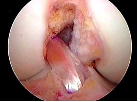
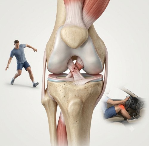
What causes ACL and PCL injuries?
Your knee has two cruciate ligaments that cross in the centre of the joint. The ACL (anterior cruciate ligament) prevents your shin bone from sliding too far forward. The PCL (posterior cruciate ligament) prevents it from sliding too far backward. Together, they provide stability when you move.
ACL injuries typically happen during sports that involve pivoting, jumping, or rapid direction changes — football, basketball, skiing, and tennis. A sudden twist or landing awkwardly can tear the ligament. Most people hear a pop and feel their knee give way immediately.
PCL injuries usually result from a direct blow to the front of the knee, such as hitting the dashboard in a car accident or falling directly onto a bent knee. Because the PCL is the strongest ligament in the knee, it takes significant force to tear it. That’s why PCL injuries are less common than ACL tears.
Signs and symptoms
ACL and PCL injuries present differently, but both affect knee stability. Recognising the signs helps determine the right course of action.
A pop at the time of injury
Many people hear or feel a pop when the ACL tears. This is less common with PCL injuries, which often result from a direct blow.
Rapid swelling
The knee swells quickly within hours of an ACL tear due to bleeding inside the joint. PCL injuries may swell more gradually.
Knee instability
The knee feels like it might give way, especially when you put weight on it or change direction.
Difficulty walking
Putting weight on the affected leg is painful, and you may need help to walk or stand.
Diagnosis
Diagnosis begins with a detailed history and physical examination. Dr. Kembhavi will perform specific tests — the Lachman test and pivot shift for ACL, and the posterior drawer test for PCL — to assess ligament integrity. An MRI confirms the diagnosis, shows the severity of the tear, and identifies any associated injuries like meniscus tears or collateral ligament damage.
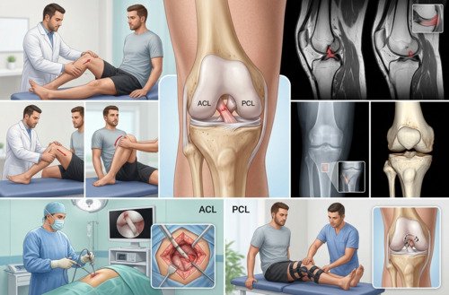
Imaging and assessment
An MRI is the gold standard for diagnosing ligament injuries. It shows whether the ligament is partially or completely torn and reveals associated injuries. X-rays are useful to rule out fractures, particularly in PCL injuries where an avulsion fracture may occur. In some cases, a CT scan may be used for surgical planning.
Treatment approach
Treatment depends on the severity of the tear, your activity level, and whether other structures are injured. For ACL tears, surgery is usually recommended for active individuals, especially those who want to return to pivoting sports. For PCL tears, many cases can be managed conservatively — the PCL has a better capacity for healing than the ACL, and isolated PCL injuries often do well with bracing and physiotherapy. Surgery is considered for high-grade tears, combined injuries, or when instability persists.
How ACL and PCL injuries are treated
Step 1
Immediate Care (PRICE)
Protection, rest, ice, compression, and elevation in the first 48–72 hours to control pain and swelling.
Step 2
Diagnostic MRI
Confirms the diagnosis, shows the severity of the tear, and identifies any associated injuries.
Step 3
Prehabilitation
Pre-operative physiotherapy to reduce swelling, restore range of motion, and strengthen the quadriceps before surgery.
Step 4
Arthroscopic Reconstruction
Keyhole surgery to replace the torn ligament with a graft, restoring stability to the knee.
Step 5
Structured Rehabilitation
A phased programme to restore range of motion, strength, and function, with clear milestones for progression.
Step 6
Return to Sport
A criterion-based return-to-sport programme, with strength and functional testing before clearance for full activity.
ACL vs. PCL treatment approaches
| Factor | ACL Reconstruction | PCL Treatment |
|---|---|---|
| Best for | Active individuals, pivoting sports, complete tears | Many isolated tears can be treated conservatively |
| Healing potential | ACL has poor blood supply and rarely heals without surgery | PCL has better healing capacity |
| Surgery threshold | Lower — surgery often recommended for return to sport | Higher — surgery for high-grade or combined injuries |
| Rehabilitation focus | Quadriceps strength is key | Strict hamstring avoidance for 4+ months |
Why choose Dr. Raghavendra Kembhavi
Arthroscopic expertise
Dr. Kembhavi performs ACL and PCL reconstruction arthroscopically through small keyhole incisions. This minimally invasive approach means less pain, less scarring, and a faster return to function compared to open surgery.
Criterion-based rehabilitation
Return to sport isn’t based on time alone — it’s based on objective strength and functional testing. Dr. Kembhavi works closely with physiotherapists to ensure you’re truly ready before clearing you for full activity.
Individualised treatment decisions
Not every ACL or PCL tear needs the same approach. Dr. Kembhavi considers your age, activity level, and goals — because a competitive athlete and a weekend walker have different needs.
Comprehensive knee assessment
Ligament injuries rarely occur in isolation. Dr. Kembhavi’s thorough diagnostic process identifies associated meniscus, cartilage, or collateral ligament injuries — ensuring they’re addressed in the same treatment plan.
Recovery timeline after reconstruction
Recovery is a marathon, not a sprint. The graft needs time to incorporate and mature before it can handle the demands of sport.
Weeks 0–6: Protection & Motion
Crutches and brace. Focus on reducing swelling, restoring range of motion, and activating the quadriceps.
Weeks 6–12: Strengthening
Progressive strengthening, balance training, and neuromuscular re-education. Walking without crutches.
Months 3–6: Advanced Training
Running, agility drills, and plyometrics introduced when strength criteria are met. Sport-specific training begins.
Months 6–12: Return to Sport
Criterion-based return to sport when strength and hop testing reach 90% of the uninvolved side.