Suprascapular Nerve Compressive Neuropathy Treatment in Bangalore
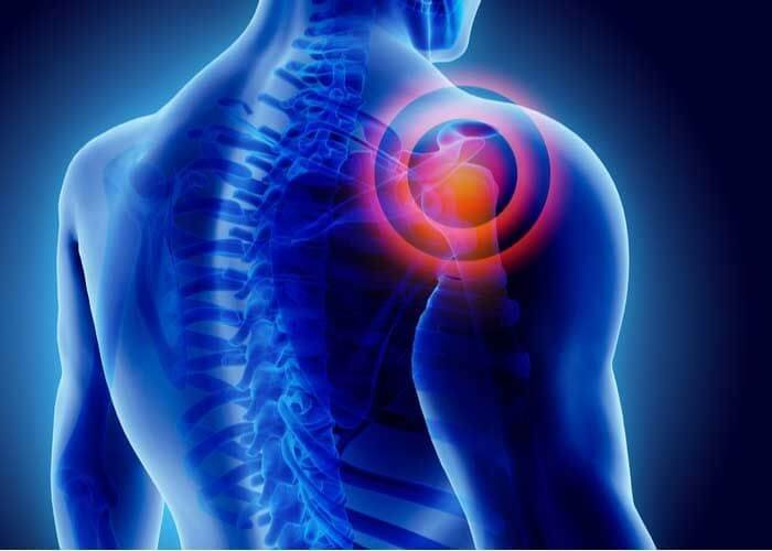
Shoulder pain that doesn’t fit the usual patterns. Weakness that develops gradually, sometimes with visible wasting of the muscles around your shoulder blade. This can be a sign of suprascapular nerve compression — a condition that’s often overlooked because it mimics more common shoulder problems.
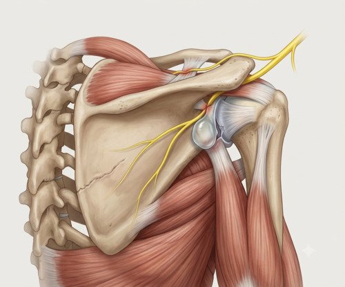
The suprascapular nerve supplies two important rotator cuff muscles — the supraspinatus and infraspinatus. When it’s compressed at the suprascapular or spinoglenoid notch, it can cause deep posterior shoulder pain, weakness with overhead activity, and eventually muscle atrophy. At our clinic in Bangalore, Dr. Raghavendra Kembhavi takes a careful diagnostic approach to identify this condition early, before irreversible nerve damage occurs.
What causes suprascapular nerve compression?
The suprascapular nerve travels from the upper brachial plexus through the posterior shoulder to supply the supraspinatus and infraspinatus muscles. Along this path, it passes through two narrow points — the suprascapular notch and the spinoglenoid notch — where it’s vulnerable to compression.
Causes include repetitive overhead activities like swimming, tennis, or volleyball, which create traction on the nerve over time. Ganglion cysts arising from labral tears are a common source of external compression. Direct trauma, shoulder dislocations, or fractures around the scapula can also injure the nerve. In some cases, no specific cause is identified.
Signs and symptoms
Suprascapular neuropathy can present in two main patterns — one dominated by pain, the other by weakness. Recognising which pattern you have helps guide treatment.
Posterior shoulder pain
Deep, aching pain behind the shoulder that’s hard to pinpoint. Often worse with overhead activity.
Weakness with overhead activity
Difficulty lifting the arm overhead or reaching behind the back. Strength may be noticeably reduced.
Muscle wasting
Visible thinning of the muscles above or below the shoulder blade. This is a late sign and indicates chronic nerve compression.
Tenderness at the notch
Pain reproduced by pressing over the suprascapular notch, or with forced cross-body abduction.
Diagnosis
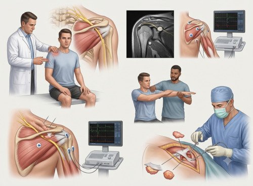
Suprascapular neuropathy is often overlooked because it mimics rotator cuff problems. Diagnosis requires a combination of clinical suspicion, imaging, and electrodiagnostic testing. Dr. Kembhavi begins with a detailed history and physical examination, looking for weakness in external rotation, tenderness at the notch, and signs of muscle atrophy.
Imaging and electrodiagnostic tests
An MRI is essential — it can show muscle edema or atrophy in the supraspinatus and infraspinatus, and identify ganglion cysts or other compressive lesions. EMG and nerve conduction studies confirm the diagnosis by showing denervation changes in the affected muscles. Together, these tests distinguish suprascapular neuropathy from rotator cuff pathology and guide treatment decisions.
Treatment approach
Treatment depends on the cause. For compression without a space-occupying lesion, non-surgical management is the first step — activity modification, physiotherapy, and anti-inflammatory medications. If a ganglion cyst or other lesion is compressing the nerve, surgical decompression is usually recommended. Arthroscopic release of the transverse scapular ligament can be performed safely and effectively, with good outcomes for pain and function.
How suprascapular neuropathy is treated
Step 1
Activity Modification
Avoiding overhead activities that aggravate the nerve. Rest from repetitive shoulder loading.
Step 2
Physiotherapy
Targeted strengthening of the rotator cuff and scapular stabilisers. Range-of-motion exercises to maintain mobility.
Step 3
Diagnostic Injection
An ultrasound-guided injection at the suprascapular notch can confirm the nerve as the pain source and provide temporary relief.
Step 4
Arthroscopic Decompression
Keyhole surgery to release the transverse scapular ligament and free the nerve. Removes any compressive cyst or lesion.
Step 5
Nerve Mobilisation
Gentle release of any adhesions around the nerve to ensure it glides freely through the notch.
Step 6
Rehabilitation
A structured programme to restore strength and function, with gradual return to overhead activity.
Surgical decompression vs. conservative treatment
| Factor | Surgical Decompression | Conservative Treatment |
|---|---|---|
| Best for | Compression from a cyst or lesion; failed conservative care | Compression without a space-occupying lesion |
| Pain relief | Direct release of the compressed nerve | Gradual; may take weeks to months |
| Strength recovery | Better outcomes when weakness is the primary symptom | Variable; may not fully restore strength |
| Recovery time | Sling 2–4 weeks; full recovery 3–6 months | No downtime; symptoms settle over 6–12 weeks |
Why choose Dr. Raghavendra Kembhavi
Accurate diagnosis matters
Suprascapular neuropathy is often mistaken for rotator cuff pathology. Dr. Kembhavi uses MRI and electrodiagnostic testing to confirm the diagnosis before recommending treatment — because the treatment pathways are completely different.
Arthroscopic expertise
When surgery is needed, Dr. Kembhavi performs arthroscopic suprascapular nerve decompression through small keyhole incisions. This approach allows direct visualisation of the nerve and precise release, with less trauma than open surgery.
A focus on nerve preservation
The goal is to relieve compression before irreversible nerve damage occurs. Early intervention can prevent permanent muscle wasting and weakness. Dr. Kembhavi prioritises timely assessment and treatment.
Individualised treatment decisions
Not every nerve compression needs surgery. Dr. Kembhavi carefully assesses each case — considering the cause, severity, and your functional goals — to determine the most appropriate treatment path.
Recovery timeline after surgical decompression
Recovery is gradual. Pain typically improves first, followed by strength as the nerve recovers.
Week 0–2
Sling for comfort. Gentle range-of-motion exercises to prevent stiffness. Pain typically begins to improve.
Week 2–6
Sling discontinued. Active range of motion. Light daily activities resume. Physiotherapy continues.
Week 6–12
Progressive strengthening begins. Return to overhead activity guided by strength recovery.
Month 3–6
Full return to sport and heavy activity for most patients. Strength continues to improve over time.