Calcific Tendinitis Treatment in Bangalore
Shoulder pain that comes on without any warning — no fall, no lifting injury, nothing you can point to as the cause. It may wake you at night, and simple movements like reaching for something or putting on a shirt suddenly feel impossible. This is often how calcific tendinitis presents, and it can be one of the most painful shoulder conditions we see.
Calcific tendinitis occurs when calcium deposits form inside a tendon — most commonly the rotator cuff in the shoulder. The pain pattern is unusual: it often peaks not when the calcium is building up, but when your body starts trying to break it down. At our clinic in Bangalore, Dr. Raghavendra Kembhavi takes a stage-based approach to diagnosing and treating this condition, because the right treatment depends entirely on where you are in the process.
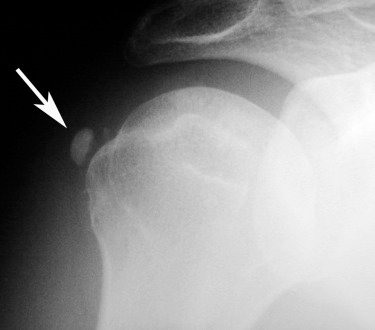
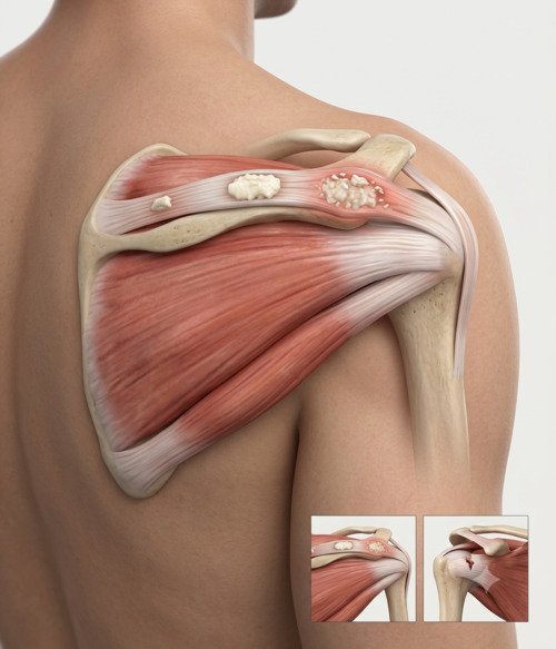
What is calcific tendinitis, and why does it hurt so much?
Calcific tendinitis is a condition where calcium crystals build up inside a tendon. It’s most common in the rotator cuff tendons of the shoulder, particularly the supraspinatus, but it can also affect the elbow, wrist, hip, and knee. It typically affects people between 30 and 60 years old and is more common in women.
What makes this condition unusual is the pain pattern. It moves through three distinct phases, and the worst pain usually doesn’t happen when the calcium is forming — it happens when your body starts breaking the deposit down. During this “resorptive” phase, the calcium softens and your immune system launches an intense inflammatory response to clear it. That’s when the severe, sometimes disabling pain hits, often waking you from sleep.
Many people mistake this pain for a rotator cuff tear, and understandably so. But the two conditions are quite different. In calcific tendinitis, the tendon fibres are usually intact — the pain comes from inflammation and pressure, not a structural gap. This distinction matters because it changes the entire treatment approach.
Signs and symptoms
The symptoms of calcific tendinitis depend on which phase the deposit is in. The pain can change dramatically over weeks or months, which can be confusing if you don’t know what’s happening.
Sudden, severe pain
Pain that comes on without warning and can be intense, often at night. It may wake you from sleep or make lying on the affected side impossible.
Difficulty moving the arm
You may be reluctant to move the shoulder at all because of the pain. Even passive movement by someone else can be uncomfortable.
Pain worse at night
Many patients describe the pain as being much worse when they’re trying to rest or sleep, which can be exhausting.
Catching or grinding sensation
Some people feel a mechanical catching or grinding when they move the shoulder, particularly in the chronic phase.
Diagnosis
Diagnosing calcific tendinitis is usually straightforward once the condition is suspected. The challenge is often that the symptoms overlap with other shoulder problems, and the pain pattern can be confusing.
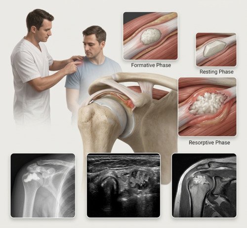
Imaging and clinical assessment
X-rays are the primary diagnostic tool — they show the calcium deposit clearly, including its size, density, and location. Interestingly, the X-ray appearance often doesn’t match the severity of symptoms; a small deposit can cause intense pain, while a large one may be relatively quiet. An ultrasound is particularly useful because it can show the stage of the deposit and whether it’s soft and fluffy (resorptive) or hard and well-defined (formative). In some cases, an MRI may be ordered to check for associated rotator cuff tears, which occur in about 30% of patients.
Understanding the stages
Calcific tendinitis moves through three phases, and knowing which phase you’re in guides the treatment. In the formative phase, calcium is being laid down — pain is usually a dull ache. In the resting phase, the deposit is stable and often painless. In the resorptive phase, your body is actively breaking the deposit down, and this is when the severe pain hits. Most people seek help during the resorptive phase, which is actually a good sign — it means the condition is starting to resolve on its own.
How calcific tendinitis is treated
Step 1
Pain Management & Rest
Anti-inflammatory medications, ice, and activity modification. Many cases settle with this alone as the deposit resolves naturally.
Step 2
Physiotherapy
Gentle range-of-motion exercises to prevent stiffness while the deposit resolves. Strengthening begins once the acute pain settles.
Step 3
Steroid Injection
A corticosteroid injection into the subacromial space can provide rapid pain relief during the acute resorptive phase.
Step 4
Barbotage (Needle Lavage)
A minimally invasive procedure where a needle breaks up and flushes out the calcium deposit under ultrasound guidance. Effective in about 60–70% of cases.
Step 5
Shock Wave Therapy
High-energy sound waves break up the deposit and stimulate healing. No incisions required. Best for chronic cases in the formative or resting phases.
Step 6
Arthroscopic Excision
For cases that don’t respond to other treatments, keyhole surgery removes the deposit directly. Reserved for a minority of patients.
Surgical vs. non-surgical treatment
| Factor | Arthroscopic Excision | Non-Surgical Treatment |
|---|---|---|
| Best for | Failed 6+ months of non-surgical care, large deposits | First-line for all patients; 90% resolve with conservative care |
| Pain relief | Direct removal of the deposit; reliable but recovery takes time | Gradual, often complete as the deposit resorbs naturally |
| Recovery time | Desk work 1–2 weeks; full recovery 3–6 months | No downtime; symptoms settle over weeks to months |
| Tendon integrity | Deposit removed; tendon left intact or repaired if needed | Tendon remains intact throughout |
Why choose Dr. Raghavendra Kembhavi
Stage-based treatment approach
Calcific tendinitis isn’t a single entity — it moves through phases, and the right treatment depends on where you are in the process. Dr. Kembhavi takes the time to identify your stage and tailor the treatment accordingly, avoiding unnecessary interventions.
A focus on natural resolution
Most cases of calcific tendinitis resolve on their own — roughly two-thirds of deposits disappear within 1–2 years. Dr. Kembhavi’s approach is to support that natural process with pain control and targeted therapy, reserving more invasive options only when needed.
Advanced arthroscopic skills
When surgery is needed, Dr. Kembhavi performs arthroscopic excision of the calcium deposit through small keyhole incisions. This allows direct removal of the deposit with minimal tissue disruption and a faster recovery than open surgery.
Accurate diagnosis matters
Calcific tendinitis is often confused with rotator cuff tears. Dr. Kembhavi uses X-ray and ultrasound together to reliably distinguish between the two — because the treatment pathways are completely different.
Recovery timeline after arthroscopic excision
Recovery from arthroscopic excision is generally quicker than from a rotator cuff repair, because the tendon itself is usually left intact. The rehab focuses on early movement and preventing stiffness.
Week 0–2: Protection & Movement
Sling for comfort only. Gentle assisted elevation and pendulum exercises begin to prevent stiffness.
Week 2–6: Active Motion
Active range of motion restored. Light daily activities resume. Return to desk work often possible by week 2–3.
Week 6–8: Strengthening
Progressive rotator cuff strengthening begins with isometrics, progressing to bands and light weights.
Month 3+: Full Recovery
Return to unrestricted activity, including overhead sports and heavy lifting, guided by strength and comfort.