Patellofemoral Instability Treatment in Bangalore
You bend your knee, and suddenly your kneecap shifts out of place. There’s a sharp pain, a feeling that the knee is giving way, and sometimes a visible deformity before it snaps back. For some, it’s a one-time event after a fall or sports injury. For others, it’s a recurring problem that makes even simple movements feel unpredictable and unsafe.
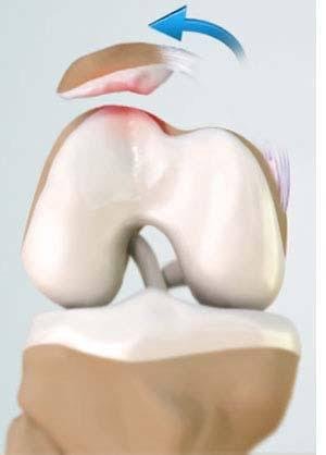
Patellofemoral instability happens when the kneecap doesn’t track smoothly within the groove at the end of the thigh bone. It can slip partially (subluxation) or completely out of place (dislocation). The condition is more common in young, active people — particularly women — and often involves underlying anatomical factors like a shallow groove, a high-riding kneecap, or malalignment. At our clinic in Bangalore, Dr. Raghavendra Kembhavi takes a comprehensive approach to patellofemoral instability, addressing both the symptoms and the underlying causes to restore stability and prevent recurrence.
What is patellofemoral instability, and why does it happen?
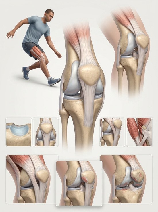
Your kneecap sits in a groove at the end of your thigh bone called the trochlea. When you bend and straighten your knee, the kneecap should glide smoothly within this groove. In patellofemoral instability, the kneecap doesn’t stay on track — it tends to slide outward, causing pain, a feeling of instability, or a complete dislocation.
The causes are often multifactorial. Structural factors include a shallow or flat trochlear groove (trochlear dysplasia), a kneecap that sits higher than normal (patella alta), or an abnormal attachment of the patellar tendon on the shin bone (increased TT-TG distance). Muscle imbalances — particularly weakness of the vastus medialis obliquus (VMO) and tightness of the lateral structures — also play a significant role. In some cases, a direct injury or loose ligaments can trigger the first episode of instability.
What’s important to understand is that instability is not just about pain. Every time the kneecap slips or dislocates, it can damage the cartilage on the underside of the kneecap and the groove. Over time, this can lead to patellofemoral osteoarthritis. This is why addressing the underlying cause — not just the symptoms — matters for long-term joint health.
Signs and symptoms
Patellofemoral instability can present in different ways. Some people have a single traumatic dislocation; others experience recurring episodes of the kneecap slipping partially out of place.
Knee pain
A dull, aching pain around the kneecap, often worse with stairs, squatting, or sitting for long periods.
A feeling of instability
The knee feels like it might give way, especially when twisting, turning, or changing direction.
Popping or cracking
A popping, grinding, or crackling sensation when you move your knee, especially with bending.
Visible dislocation
In a complete dislocation, the kneecap visibly pops out of place, usually to the outside of the knee, before returning.
Diagnosis
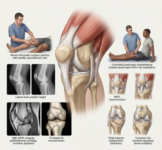
Diagnosis begins with a detailed history and physical examination. Dr. Kembhavi will assess your knee for the “apprehension sign” — a feeling of anxiety when the kneecap is gently pushed outward, which suggests instability. Imaging is essential to identify the underlying anatomical factors that contribute to the problem.
Imaging and assessment
X-rays assess the patellar height and alignment. MRI is the imaging study of choice — it shows the condition of the cartilage, the integrity of the medial patellofemoral ligament (MPFL), and the presence of trochlear dysplasia or patella alta. CT scans are useful for measuring the TT-TG distance (tibial tuberosity-trochlear groove), which helps determine whether a tibial tubercle osteotomy is needed. Together, these imaging studies provide a complete picture of the anatomical factors contributing to your instability.
Treatment approach
Non-surgical treatment is the first step for many patients, especially those with a first-time dislocation and no significant anatomical abnormalities. Physiotherapy focuses on strengthening the quadriceps — particularly the VMO — and the hip stabilisers to improve patellar tracking. Surgical treatment is considered for recurrent instability, significant anatomical risk factors, or when conservative treatment fails. The goal is to address the underlying cause — whether that’s reconstructing the MPFL, realigning the tibial tubercle, or correcting trochlear dysplasia.
How patellofemoral instability is treated
Step 1
Physiotherapy
Strengthening the quadriceps (especially VMO) and hip stabilisers. Gait and proprioceptive training. The first-line treatment for most cases.
Step 2
MPFL Reconstruction
The medial patellofemoral ligament — which stabilises the kneecap on the inner side — is reconstructed using a tendon graft. This is the cornerstone of surgical treatment.
Step 3
Tibial Tubercle Osteotomy
When malalignment is present, the attachment of the patellar tendon is moved to improve patellar tracking. May be performed as a distalization or medialization.
Step 4
Trochleoplasty (Selected Cases)
For severe trochlear dysplasia, the groove is reshaped to provide a deeper track for the kneecap. Reserved for high-grade dysplasia.
Step 5
Structured Rehabilitation
A phased programme that protects the reconstruction while restoring range of motion and strength, with clear milestones for progression.
Step 6
Return to Sport
Gradual return to sport guided by strength testing and functional readiness. Full clearance typically 7–12 months after surgery.
Non-Surgical vs. Surgical Treatment
| Factor | Non-Surgical | Surgical |
|---|---|---|
| Best for | First-time dislocation, no major anatomical risk factors | Recurrent instability, significant anatomical factors, failed conservative care |
| Approach | Physiotherapy, bracing, gait training | MPFL reconstruction, tibial tubercle osteotomy, trochleoplasty |
| Recovery time | No downtime; 6–12 weeks of guided rehab | Brace 6 weeks; full recovery 7–12 months |
| Recurrence risk | Higher in patients with anatomical risk factors | Lower; addresses the underlying cause |
Why choose Dr. Raghavendra Kembhavi
Comprehensive assessment of all risk factors
Patellofemoral instability is multifactorial. Dr. Kembhavi assesses all contributing factors — trochlear morphology, patellar height, alignment, and muscle balance — to create a personalised treatment plan that addresses the root cause, not just the symptoms.
Arthroscopic and minimally invasive techniques
When surgery is needed, Dr. Kembhavi uses arthroscopic and minimally invasive techniques — MPFL reconstruction, tibial tubercle osteotomy, and trochleoplasty — to restore stability with less trauma and faster recovery.
Focus on long-term joint preservation
Every instability episode can damage the cartilage. Dr. Kembhavi’s approach focuses on restoring stability to protect the joint and prevent the progression to patellofemoral osteoarthritis.
Structured rehabilitation
Rehabilitation is critical to a good outcome. Dr. Kembhavi works closely with physiotherapists to ensure a structured, phased programme that protects the reconstruction and restores function safely.
Recovery timeline after MPFL reconstruction
Recovery is phased and requires patience. The reconstructed ligament needs time to heal, and progression is based on achieving specific milestones.
Weeks 0–2: Protection
Crutches and brace. Gentle range of motion. Focus on reducing swelling and protecting the reconstruction.
Weeks 3–6: Early Rehab
Progressive range of motion and quadriceps activation. Brace remains for walking until quad control is adequate.
Weeks 7–12: Strengthening
Progressive strengthening, balance drills, and low-impact activities. No running or jumping yet.
Months 3–7: Return to Sport
Functional and sport-specific training. Return to sport typically around 7 months. Approximately 90% return to sport.