Medial Collateral Ligament (MCL) Injury Treatment in Bangalore
You felt it on the inside of your knee — a sharp pain after a twist, a collision, or a sudden sideways force. Within hours, the knee started to swell, and putting weight on it felt unstable. This is often how an MCL injury presents.
The medial collateral ligament (MCL) is one of the four major ligaments stabilising your knee. It runs along the inner side of the joint, connecting the thigh bone to the shin bone, and resists forces that push the knee inward. MCL injuries are common in sports like football, skiing, and rugby, and they account for a significant proportion of all knee ligament injuries. The good news is that most MCL injuries heal well without surgery. At our clinic in Bangalore, Dr. Raghavendra Kembhavi takes a graded approach — treating each injury according to its severity and only recommending surgery when it’s genuinely needed.
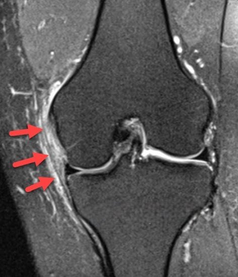
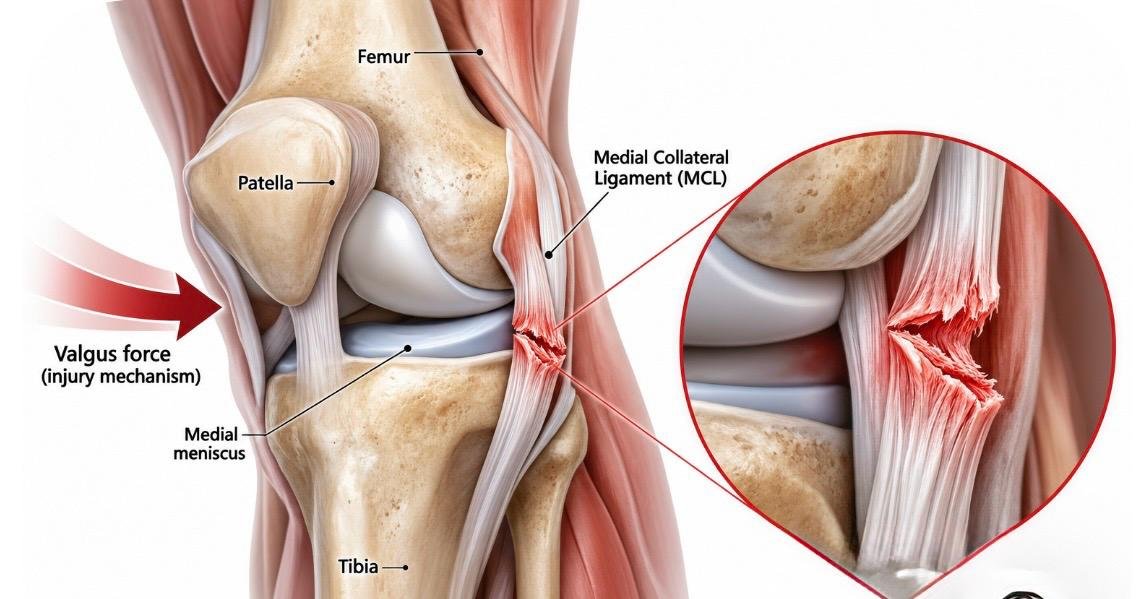
What is the MCL, and how do injuries happen?
The MCL is a strong band of tissue on the inner side of your knee. It has two parts — a superficial layer and a deeper layer — and it connects the femur (thigh bone) to the tibia (shin bone). Its main job is to resist valgus stress — forces that push the knee inward .
Most MCL injuries occur when a force hits the outside of the knee, pushing it inward. This can happen during a tackle in football, a fall while skiing, or a collision in rugby. The ligament can also be injured from a twisting motion or by landing awkwardly. In many cases, MCL injuries occur alongside other knee injuries, particularly ACL tears — this combination is sometimes called the “unhappy triad” .
MCL injuries are graded from I to III. Grade I is a mild sprain with microscopic tearing. Grade II is a partial tear with some laxity. Grade III is a complete tear with significant instability. The grade determines the treatment approach, but the good news is that the MCL has a good blood supply and most injuries — even complete tears — heal well without surgery.
Signs and symptoms
The symptoms of an MCL injury depend on the grade, but the pattern is fairly characteristic. The pain is almost always on the inner side of the knee.
Pain on the inner knee
Sharp or aching pain along the inner side of the knee, often made worse by pushing the knee inward.
Swelling
Swelling develops within hours to days, depending on the severity. Grade III tears often produce more swelling.
Instability
The knee may feel like it’s giving way, especially with side-to-side movements or when pivoting.
Difficulty walking
Putting weight on the affected leg may be painful, especially with higher-grade injuries. You may need crutches initially.
Diagnosis
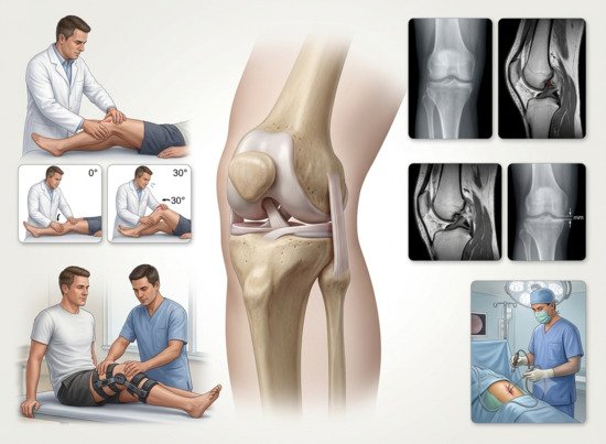
Diagnosis begins with a detailed history — how the injury happened, what movements bring on the pain, and whether there was any swelling or instability. Dr. Kembhavi performs a careful physical examination, including the valgus stress test at 0° and 30° of knee flexion. This test reproduces the pain and assesses the degree of laxity, which helps determine the grade of injury.
Imaging and assessment
X-rays are usually normal in MCL injuries but help rule out fractures. MRI is the imaging study of choice — it confirms the grade of injury and identifies any associated injuries, particularly ACL or meniscal tears, which occur in a significant number of cases . Stress radiographs may be used in some cases to quantify the degree of laxity.
Treatment approach
The treatment of MCL injuries is graded. Grade I and II injuries are almost always treated non-surgically — rest, bracing, and physiotherapy. Grade III (complete) tears in isolation also heal well without surgery in most cases, thanks to the MCL’s good blood supply. Surgery is considered for Grade III tears associated with other ligament injuries (like ACL tears), for chronic instability, or when the MCL fails to heal with conservative treatment.
How MCL injuries are treated
Step 1
Immediate Care (PRICE)
Protection, rest, ice, compression, and elevation in the first 48–72 hours to reduce pain and swelling.
Step 2
Bracing
A hinged knee brace protects the healing ligament while allowing controlled motion. Braces are typically worn for 4–6 weeks.
Step 3
Physiotherapy
The cornerstone of MCL treatment. Range-of-motion exercises, quadriceps and hamstring strengthening, and progressive return to activity.
Step 4
Surgical Repair (If Needed)
Reserved for Grade III tears with associated ACL injury, chronic instability, or failed conservative care. The MCL is repaired or reconstructed.
Step 5
Structured Rehabilitation
A phased programme that progresses from protected motion to full activity, with clear criteria for advancement.
Step 6
Return to Sport
Gradual return to sport guided by strength testing and functional readiness. Full clearance is based on objective criteria, not just time.
Non-surgical vs. surgical treatment
| Factor | Non-Surgical | Surgical |
|---|---|---|
| Best for | Grade I–III isolated tears; most MCL injuries | Grade III with ACL injury, chronic instability, failed conservative care |
| Healing potential | MCL has good blood supply; most heal well | Direct repair or reconstruction for severe cases |
| Recovery time | Grade I: 1–2 weeks; Grade II: 3–6 weeks; Grade III: 6–12 weeks | Bracing and protected weight-bearing for 6 weeks; full recovery 3–6 months |
| Return to sport | Guided by grade and functional testing | Criterion-based, typically 4–6 months |
Why choose Dr. Raghavendra Kembhavi
Most MCL injuries don’t need surgery
Unlike ACL injuries, most MCL tears heal well without surgery — even complete tears. Dr. Kembhavi takes a graded approach, reserving surgery for the small percentage of cases that genuinely need it.
Comprehensive knee assessment
MCL injuries often occur alongside ACL or meniscal tears. Dr. Kembhavi’s thorough diagnostic process identifies associated injuries, ensuring they’re addressed in the same treatment plan.
Structured rehabilitation
Rehabilitation is the cornerstone of MCL treatment. Dr. Kembhavi works closely with physiotherapists to ensure a structured programme that progresses from protected motion to full activity, with clear milestones for progression.
Criterion-based return to sport
Return to sport isn’t based on time alone — it’s based on objective strength and functional testing. This reduces the risk of re-injury and ensures you’re truly ready before returning to full activity.
Recovery timeline for MCL injuries
Recovery depends on the grade of injury. The good news is that most MCL injuries heal without surgery, and the timeline is predictable.
Week 0–2: Protection
Brace and crutches as needed. Ice and elevation. Gentle range of motion exercises begin within days.
Week 2–6: Motion & Strengthening
Progressive range of motion and strengthening. Crutches discontinued for most. Bracing continues for Grade II–III injuries.
Week 6–12: Functional Training
Advanced strengthening, balance, and sport-specific drills. Bracing typically discontinued by week 6–8 for Grade I–II injuries.
Month 3+: Return to Sport
Return to sport guided by functional testing. Grade I–II injuries typically return by 3–6 weeks; Grade III by 3 months or more.