Shoulder Dislocation Treatment in Bangalore for Stability, Recovery & Return to Activity
A shoulder dislocation occurs when the upper end of the arm bone moves out of its normal position within the shoulder socket. It is a common injury that can occur during sports, falls, accidents or sudden impact and may cause severe pain, swelling and difficulty moving the arm.
Shoulder dislocation treatment in Bangalore focuses on restoring the shoulder to its proper position, assessing any associated damage and reducing the risk of recurrent instability. Treatment depends on the type of dislocation, the patient’s age, activity level, previous dislocations and the condition of the surrounding bones, ligaments, labrum and tendons.
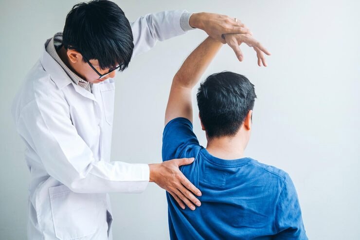

What Is a Shoulder Dislocation?
The shoulder is one of the body’s most mobile joints, but this mobility also makes it more susceptible to instability and dislocation.
A shoulder dislocation happens when the head of the upper arm bone comes out of the socket. Most dislocations occur towards the front of the shoulder and are known as anterior shoulder dislocations, although posterior and other types can also occur.
A dislocation can stretch or damage the ligaments and labrum around the shoulder and, in some cases, may be associated with bone or tendon injuries.
Types of shoulder dislocation
The direction and pattern of dislocation, along with how many times it has happened, shape the right treatment approach.
Anterior Dislocation
Most Common
The arm bone slips forward out of the socket — accounts for the large majority of shoulder dislocations, typically from a fall or forced overhead position.
Posterior Dislocation
Less Common
The arm bone slips backward, often from a direct blow to the front of the shoulder, a seizure, or an electric shock injury.
First-Time (Acute) Dislocation
By Frequency
A single dislocation event, often from a specific injury, treated with reduction and a structured rehabilitation program.
Recurrent Instability
By Frequency
Repeated dislocation or a persistent feeling of the shoulder “giving way,” usually needing surgical stabilisation to restore lasting stability.
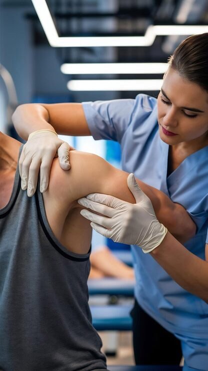
First-Time vs Recurrent Shoulder Dislocation
The treatment approach can be different after a first-time dislocation compared with recurrent instability.
First-Time Dislocation
After the shoulder is reduced, the joint is assessed for associated injuries. Treatment may include immobilisation followed by structured physiotherapy and rehabilitation.
Recurrent Dislocation
If the shoulder repeatedly comes out of position, there may be underlying damage to the labrum, ligaments or bone. In active patients or those with recurrent instability, shoulder stabilisation surgery may be considered.
The decision depends on the patient’s age, sports participation, number of dislocations, structural damage and functional requirements.
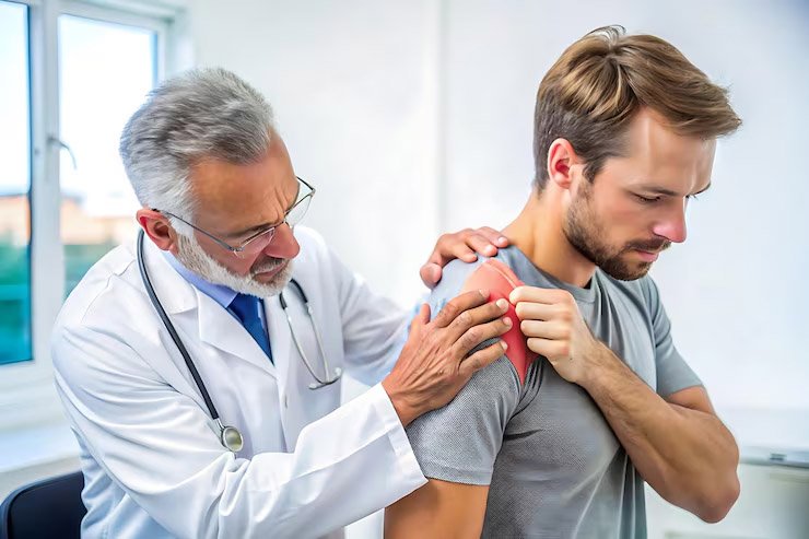
Signs of an acute shoulder dislocation
These signs suggest a dislocation has occurred and warrant urgent evaluation.
- Visibly deformed or out-of-place shoulder
- Severe pain immediately after the injury
- Inability to move the arm
- Swelling or bruising around the shoulder
- Numbness, tingling or weakness down the arm
- A feeling of the shoulder being “out of the socket”
- Muscle spasm around the shoulder joint
- For recurrent instability: repeated episodes or a sense the shoulder may “give way” during activity
How shoulder dislocation is treated, step by step
Step 1
Emergency Reduction
A controlled, urgent procedure to guide the shoulder back into its socket, usually with sedation or pain relief for comfort.
Step 2
Imaging
X-ray to confirm successful reduction and rule out a fracture; MRI to assess labral or ligament damage.
Step 3
Immobilisation
A sling protects the shoulder for 1 to 3 weeks while initial healing begins.
Step 4
Physiotherapy
Progressive rehabilitation restores motion and rebuilds strength in the muscles that support shoulder stability.
Step 5
Risk Assessment
Your age, activity level, sport, and any structural damage on imaging are reviewed to assess the risk of further dislocation.
Step 6
Stabilisation Surgery (If Needed)
For recurrent instability or high re-dislocation risk, arthroscopic surgery restores the joint’s natural stability.
Non-surgical rehabilitation vs. stabilisation surgery
| Factor | Non-Surgical Rehabilitation | Arthroscopic Stabilisation Surgery |
|---|---|---|
| Best for | First-time dislocation, lower-risk patients | Recurrent dislocation, young athletes, high-risk activity |
| Re-dislocation risk | Higher, especially in young, active patients | Significantly reduced with successful repair |
| Recovery | 6–12 weeks of physiotherapy | 3–4 weeks in a sling, several months of rehab |
| Return to contact sport | Variable, with ongoing instability risk | 5–6 months, with restored stability |
Arthroscopic Shoulder stabilisation surgery
When the shoulder continues to dislocate or feel unstable, surgery restores the structures that keep the joint in place. Dr. Raghavendra Kembhavi performs advanced leading arthroscopic stabilisation techniques, selection based on the extent of damage found on imaging.
Arthroscopic Bankart Repair
Minimally invasive; performed using a camera (arthroscope) through small incisions
The torn labrum and capsule are reattached to the glenoid rim using suture anchors
Ideal for patients with soft tissue injury and minimal bone loss
Remplissage Procedure
Performed in conjunction with Bankart repair
Fills a Hill-Sachs defect (compression fracture of the humeral head) with the infraspinatus tendon
Prevents engagement of the defect with the glenoid rim
Capsular Shift or Plication
Tightens the loose joint capsuleOften combined with other procedures
A bone block procedure
This novel procedure is performed arthroscopically for recurrent dislocation or instability of the shoulder with subcritical bone loss (10-15%) in the glenoid bone. This procedure utilizes a bone graft ideally taken from clavicle end, spine of scapula or pelvic bone to address and fill the defect either in front (anterior bone block) or back (posterior bone block) of glenoid bone. The graft will be secured to the defect in the glenoid with screws or sutures.
Latarjet Procedure
Open surgery (can also be done arthroscopically by experienced surgeons). A portion of the coracoid process (a bony projection from the scapula) is transferred to the front of the glenoid. Provides both bone support and a “sling effect” from the muscle attached to the coracoid. Preferred for patients with significant bone loss or failed prior stabilization surgery.
A detailed recovery timeline
The pathway differs depending on whether surgery is needed — both are outlined below.
Day 0: Reduction & Immediate Care
The shoulder is relocated under sedation or pain relief; an X-ray confirms successful reduction and checks for fracture.
Week 1–3: Sling & Protection
The arm is protected in a sling; gentle pendulum exercises may begin early to prevent stiffness.
Week 8–12: Functional Return
Most patients resume daily activities and low-demand work; sport-specific training begins where appropriate.
If Surgery Is Needed — Week 0–4
Arthroscopic Bankart repair or Latarjet procedure performed; the arm is protected in a sling during initial healing.
If Surgery Is Needed — Month 2–4
Structured physiotherapy restores motion and progressively rebuilds strength around the repaired structures.
If Surgery Is Needed — Month 5–6
Return to full activity and contact sport, guided by strength testing and functional readiness.
Fellowship-trained, sports medicine focused
Dr. Raghavendra Kembhavi is a fellowship-trained Orthopaedic Surgeon and Sports Medicine Specialist offering minimally invasive arthroscopic care for hip, knee and shoulder conditions in Bangalore.

10000+
Successful Patient Consultations

3000+
Orthopaedic & Sports Injury Treatments

2500+
Arthroscopic & Joint Procedures

17+
Years of Trusted Experience